
Page 71 - eBook_Proceedings of the International Conference on Digital Manufacturing V2
P. 71
Fabrication and Characterization of a Low-Cost Piezoelectric using Rochelle
Salt for Energy Harvesting and Sensor Applications
Complementing these metrics, qualitative results (Figure 27)
showcase visual segmentation outputs that demonstrate the
model’s ability to accurately delineate and classify cervical cell
regions within Pap smear images.
Together, these results affirm the model’s potential for reliable
classification of normal, precancerous, and cancerous cells,
contributing meaningfully to cervical cancer screening
applications.
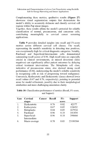
Table 9 provides detailed insights into recall and F1-score
metrics across different cervical cell classes. The recall,
representing the model's sensitivity in detecting true positives,
was exceptionally high for critical diagnostic categories. Notably,
Parabasal and Superficial-Intermediate cells demonstrated
outstanding recall scores of 0.96. High recall rates are especially
crucial in clinical environments, as missed detections (false
negatives) can significantly affect patient outcomes by delaying
critical treatment interventions. The Metaplastic cell class,
indicative of precancerous states, also showed strong recall
performance (0.84), underscoring the model’s reliable sensitivity
in recognising cells at risk of progressing toward malignancy.
Conversely, Koilocytotic and Dyskeratotic classes showed lower
recall values (0.67 and 0.76, respectively), pointing to potential
areas for model refinement, possibly due to their morphological
similarities and more challenging annotation clarity.
Table 10: Classification performance of metrics (Recall, F1-score,
and Support)
S.no Cervical Cancer Recall F1- Support
stages score
1. Dyskeratotic 0.76 0.02 42177
2. Koilocytotic 0.67 0.05 124452
3. Metaplastic 0.84 0.05 167934
4. Parabasal 0.96 0.10 141854
5. Superficial- 0.96 0.10 398464
Intermediate
6. Macro average 0.70 0.05 23189504
55